NAIL ANATOMY – Different Parts of Fingernail
Nail Anatomy
Authors: Doug Schoon and Ana Seidel
Nail Anatomy – The Different Parts of the Fingernail
Do you know where your cuticle is? Or your hyponychium? Most people don’t.
Not only is the general public confused about the names for the parts of the natural nail, but many nail technicians are not able to name the various major parts and know their function.
Let’s change this today.
Matrix
Where new nail plate cells are created and the nail plate begins to form.
Lunula
Lunula
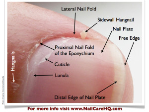
A bluish-white, opaque area that is visible through the nail plate. This area is the front part of the nail matrix. Sometimes, it’s called the “moon.”
The lunula is the front part of the matrix we can see, or in other words, the visible matrix.
Not all fingers have a visible lunula. Usually, it is easiest to find a lunula on a thumb or index finger.
Many people think that they would like to have lunula’s, but in fact, you really don’t.
Since it is the exposed portion of the matrix, this area is not protected by the eponychium. It is easy bruised with every day life tasks.
Those bruises show up as little white marks in the nail plate.
Eponychium
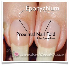
Living skin at the base of the nail plate that covers the matrix area. This should NOT be confused with the “cuticle”.
Proximal Fold of the Eponychium
Healthy Proximal Fold
A tight band of living tissue that most people incorrectly think is their “cuticle”.
Since this skin dries out easily, people are quick to clip this skin believing that it’s not necessary.
Please, please, please…DO NOT CUT THIS SKIN!
The proximal fold is a required guardian seal that prevents germs and bacteria from getting to the nail matrix, where new cells are created.
I always know when girls are cutting. Their entire cuticle line is red and inflamed. Basically, their eponychium is infected all the time.
If you go to a salon for a manicure, do not ever let your nail tech cut this skin.
The best way to keep this skin soft and tight to the nail plate is with a high quality, jojoba wax ester based penetrating nail and cuticle oil.
Cuticle
*Represented as a pink line in the first photo
CLICK PICTURE FOR LARGER DETAIL
A thin layer of dead tissue riding on the nail plate to form a seal between the nail plate and eponychium to prevent pathogens from infecting the matrix area.
The cuticle pulls away from the underside of the eponychium and attaches tenaciously to the nail plate.
The cuticle should NOT be confused with the “eponychium”.
Since polish and nail enhancements don’t bond to skin on the nail plate, the cuticle should be properly removed with gentle scraping only.
Nippers can’t remove the cuticle. It’s physically impossible.
Nail Plate
Composed of hardened, flat, translucent, non-living, keratin nail cells that form a solid, protective layer over the underlying soft tissue.
The nail plate should NOT be confused with the nail “bed”.
The average person has 50 layers of keratin cells that make up the nail plate.
The thickness of your nails is determined by the size of your matrix.
Not everyone’s matrix is the same size. People with thin nails have a small matrix and will have less than 50 layers. People with thick nails have a large matrix and have more than 50 layers.
Nail Bed
The soft, pink tissue that sits underneath and supports the nail plate while it grows. The nail bed should NOT be confused with the nail “plate”.
CLICK FOR MORE DETAIL
Hyponychium
*Represented as a pink line in the first photo
A soft tissue seal underneath the extended “free” edge of the nail plate whose purpose is to prevent pathogens from infecting the nail bed.
Onychodermal Band
A band of bunched up tissue located behind the hyponychium.
This band improves the ability of the hyponychium to prevent pathogens from infecting the nail bed.
The onychodermal band works in the same way as the proximal fold on the top surface of the nail.
When looking at your bare nails, you can see this darker band of skin right before your nail plate leaves the nail bed to become your free edge.
Solehorn Cuticle
A thin layer of dead tissue riding on the nail plate to form a seal between the nail plate and hyponychium to prevent pathogens from infecting the nail bed.
The solehorn cuticle pulls away from the underside of the hyponychium and attaches tenaciously to the nail plate.
Bone
Bone supports and shapes both the nail matrix and nail bed. The flat or curved shape of your nails is determined by the shape of your fingertip bone.
Knowledge is Power
Knowing your nail anatomy is important for the home manicurist and can actually help you find an excellent nail professional if you’re wanting to be pampered.
Ask her how she removes the cuticle.
If she shows you nippers instead of a spoon shaped remover or curette, you’ll know to politely walk out and find someone new.
Remove Your Cuticles the RIGHT Way for Healthy Nails
- It is extremely dangerous to cut proximal folds (cuticles) because they protect the nail matrix where nail cells are created.
- Cuticle cutting, removing dead or live skin with a drill is also very dangerous.
- AVOID from Dry Manicures/Russian Manicures.
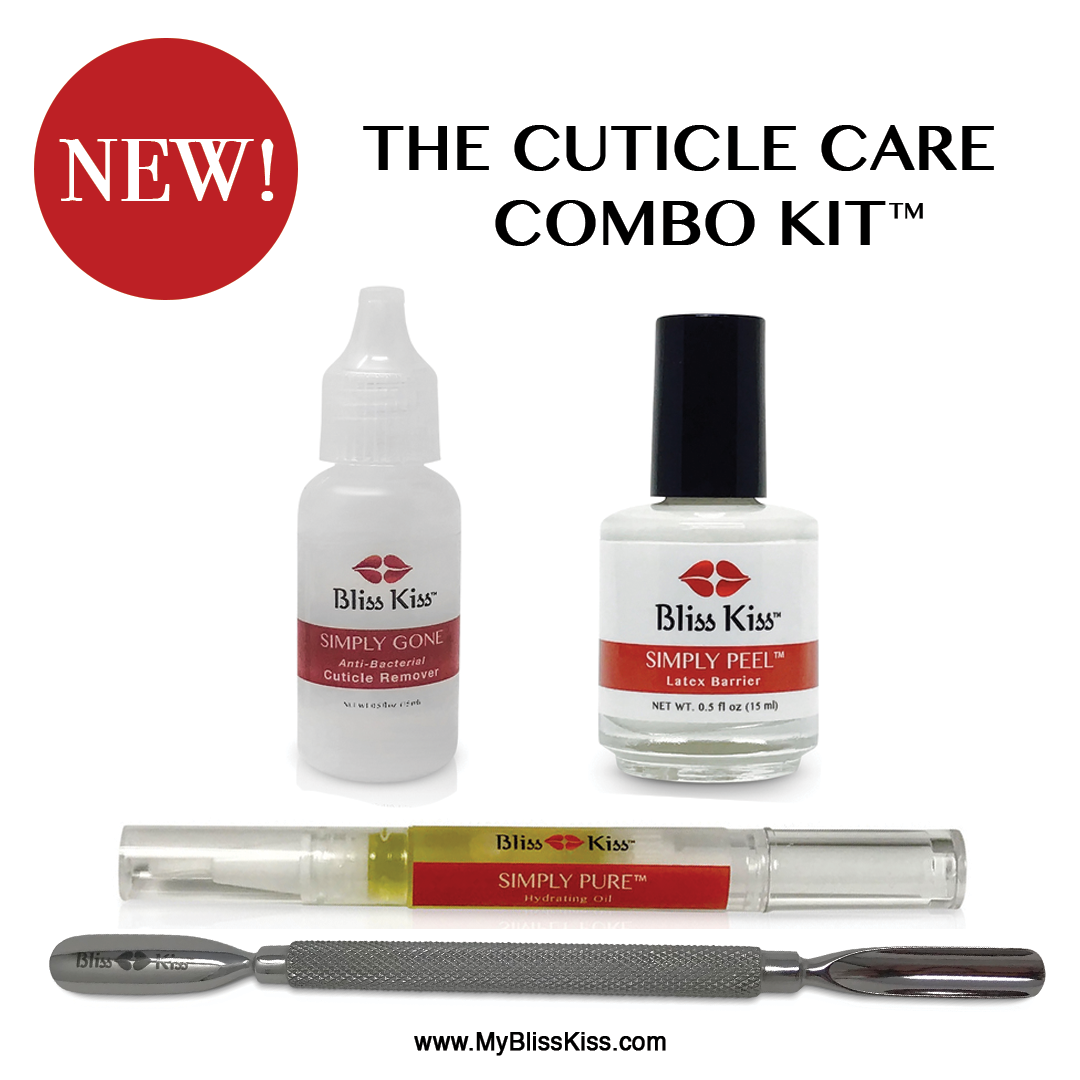
The dead cuticle skin needs to be removed with the correct tools.
